Human & Animal Primary Cells: Physiologically Relevant Models for Modern Life-Science Research

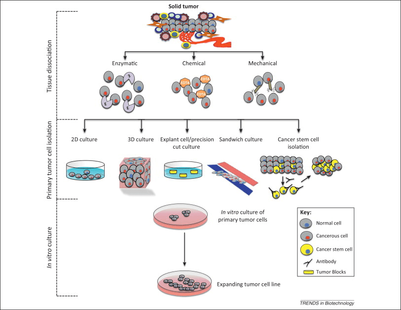
Human & animal primary cells are cells isolated directly from living tissues or blood and cultured for a limited lifespan under controlled laboratory conditions. Because they have not undergone immortalization or extensive genetic manipulation, primary cells preserve native morphology, signaling pathways, metabolic states, and functional responses, making them indispensable tools for physiologically relevant in vitro research.

Gene Knockdown Stable Cell Lines: An In-Depth Educational Resource for Laboratory Research

Introduction

Gene Knockdown Stable Cell Lines are a cornerstone of modern molecular and cellular biology. They enable researchers to reduce gene expression in a stable, heritable, and reproducible manner, making them ideal for long-term experiments, pathway analysis, and mechanistic studies. Because gene knockdown does not completely eliminate gene function, these systems are especially valuable for studying essential genes, regulatory networks, and dosage-dependent biological effects.

Extensive educational background on gene regulation, molecular genetics, and experimental models is available from government and academic institutions such as https://www.nih.gov, https://www.ncbi.nlm.nih.gov, and https://www.genome.gov.

Understanding Gene Knockdown Versus Gene Knockout

Gene knockdown refers to partial suppression of gene expression, typically at the mRNA or transcriptional level, whereas gene knockout results in complete loss of gene function. In laboratory research, knockdown approaches often provide more physiologically relevant insights, particularly when total gene loss leads to lethality or compensatory artifacts.

Introductory and advanced explanations of gene expression control can be found at:

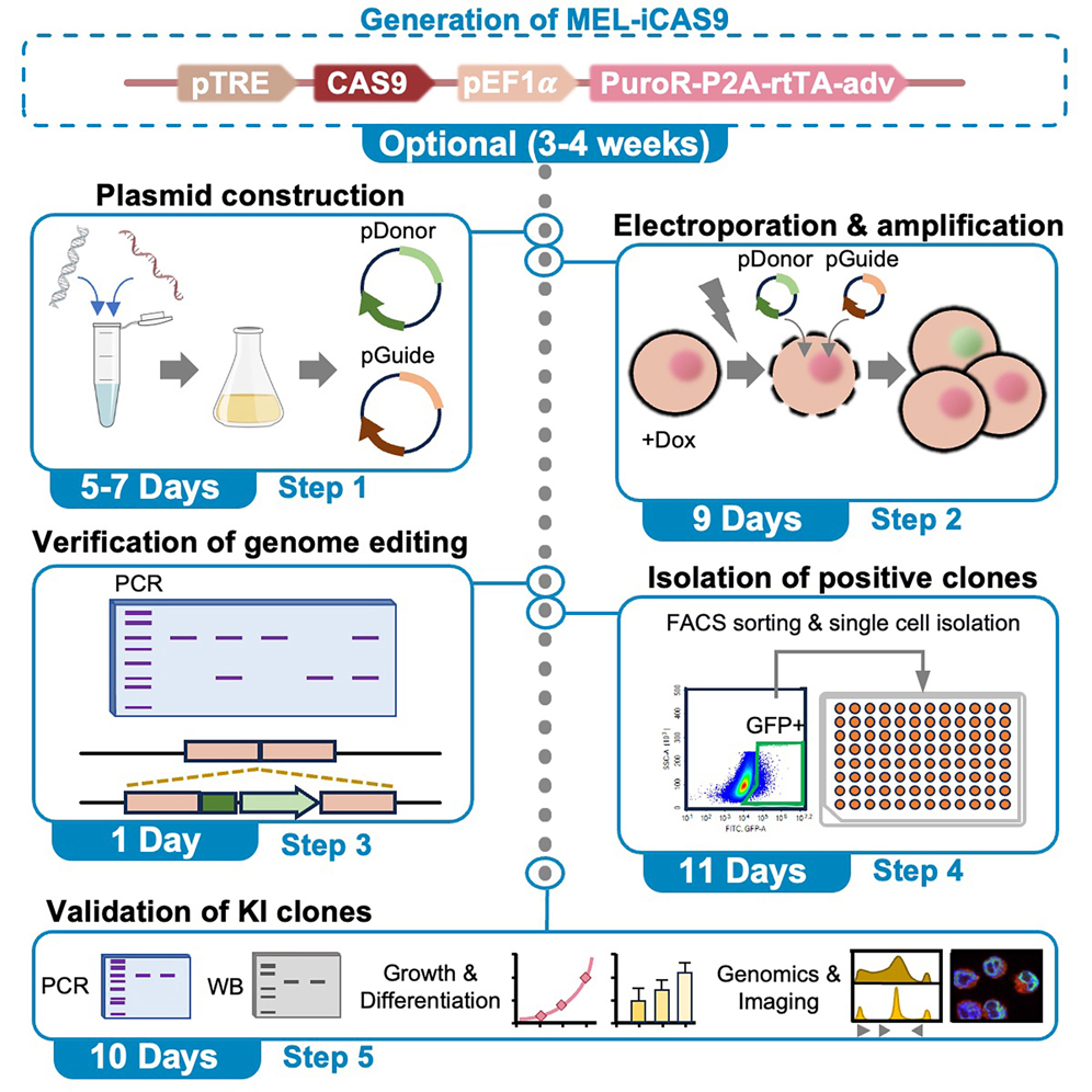
Protocol for efficient CRISPR-Cas9-mediated fluorescent tag ...

Technologies Used to Generate Stable Gene Knockdown Cell Lines

RNA Interference (RNAi) and shRNA

Short hairpin RNA (shRNA)–based knockdown is one of the most widely adopted methods for generating stable gene knockdown cell lines. shRNA constructs are commonly delivered using lentiviral vectors, allowing genomic integration and long-term expression. Once transcribed, shRNA is processed into siRNA by the endogenous RNAi machinery, resulting in degradation of the target mRNA.

Authoritative resources explaining RNA interference include:

CRISPR Interference (CRISPRi)

CRISPR interference (CRISPRi) relies on a catalytically inactive Cas9 protein (dCas9) fused to transcriptional repressors. This system binds specific genomic loci and blocks transcription without inducing DNA breaks. CRISPRi provides high specificity, tunable repression, and reversibility, making it increasingly popular in functional genomics.

Educational and policy-level information on CRISPR technologies is available at:

Workflow for Establishing Gene Knockdown Stable Cell Lines

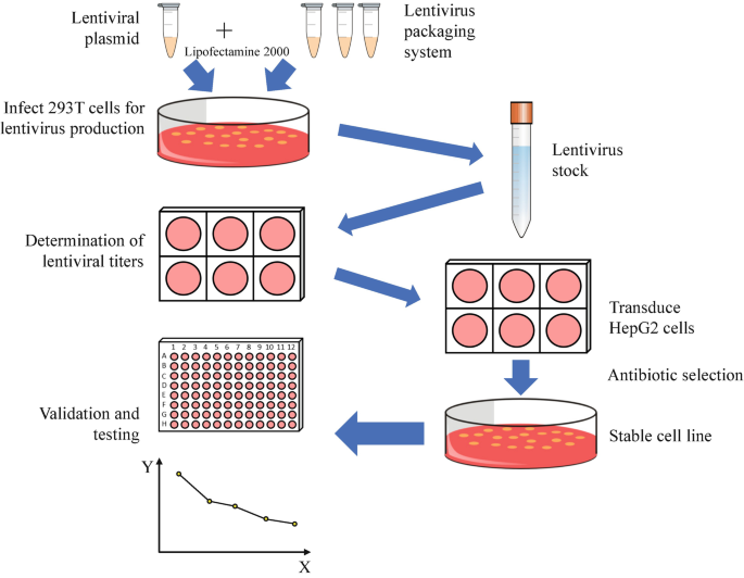
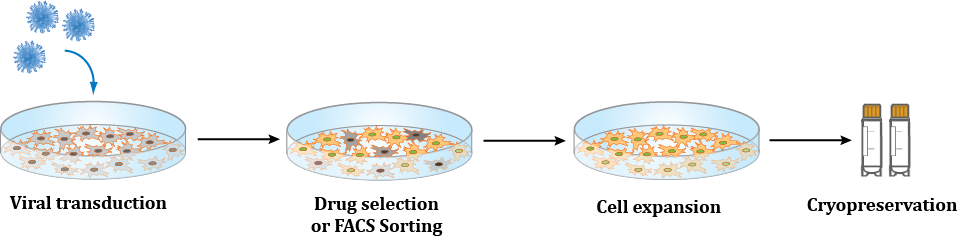
A typical laboratory workflow includes:

  1. Design of shRNA or guide RNA targeting the gene of interest

  2. Vector construction and viral packaging

  3. Cell transduction or transfection

  4. Antibiotic or fluorescence-based selection

  5. Validation of knockdown efficiency

  6. Functional and phenotypic characterization

Detailed laboratory workflows and best practices are documented at:

Analyzing efficiency of a lentiviral shRNA knockdown system in ...

Key Laboratory Applications

Functional Genomics

Stable gene knockdown cell lines enable systematic interrogation of gene function across biological pathways, supporting transcriptomic, proteomic, and phenotypic analyses.

Cancer Biology and Disease Modeling

By stably suppressing oncogenes, tumor suppressors, or disease-associated genes, researchers can model disease mechanisms and investigate cellular dependencies.

Drug Discovery and Target Validation

Gene knockdown stable cell lines are widely used to validate therapeutic targets and assess pathway sensitivity in preclinical research.

Signal Transduction and Network Biology

Reducing expression of receptors, kinases, or transcription factors allows detailed mapping of signaling cascades and regulatory networks.

Methods for Establishing and Using a Stable Cell Line Expressing ...

Advantages of Gene Knockdown Stable Cell Lines

  • Long-term, consistent gene suppression

  • Improved reproducibility across experiments

  • Reduced variability compared to transient methods

  • Compatibility with high-throughput and long-term studies

  • Enhanced relevance for essential gene research

Guidelines on experimental rigor and reproducibility are provided by:

Validation and Quality Control Strategies

Validation is critical to ensure biological relevance. Common approaches include:

  • Quantitative PCR for transcript analysis

  • Western blot or immunocytochemistry for protein detection

  • Functional assays aligned with gene function

Reference standards and validation guidance:

Emerging and Advanced Research Applications

Gene knockdown stable cell lines are increasingly applied in:

Conclusion

Gene Knockdown Stable Cell Lines are essential experimental systems for long-term, reproducible analysis of gene function. Their ability to model partial gene suppression makes them uniquely suited for functional genomics, disease research, and drug discovery. By combining robust molecular tools with rigorous validation, these cell lines continue to advance laboratory research across a wide range of scientific disciplines.

Signaling Pathway Reporter Cell Lines in Laboratory Research: Quantitative Pathway Analysis, Functional Readouts, and Assay Development

Introduction: Why Signaling Pathway Reporter Cell Lines Are Foundational Research Tools

Signaling Pathway Reporter Cell Lines are genetically engineered cell lines designed to translate intracellular pathway activation into a quantifiable reporter signal, such as luminescence or fluorescence. These systems allow researchers to directly monitor pathway dynamics in living cells with high sensitivity and reproducibility.

According to the National Center for Biotechnology Information, cellular signaling pathways regulate gene expression, metabolism, proliferation, differentiation, and stress responses
https://www.ncbi.nlm.nih.gov/books/NBK26878/

In laboratory research, pathway reporter cell lines are indispensable for:

  • Functional pathway mapping

  • Mechanism-of-action studies

  • Drug and compound screening

  • Toxicology and stress response modeling

  • Signal transduction kinetics analysis

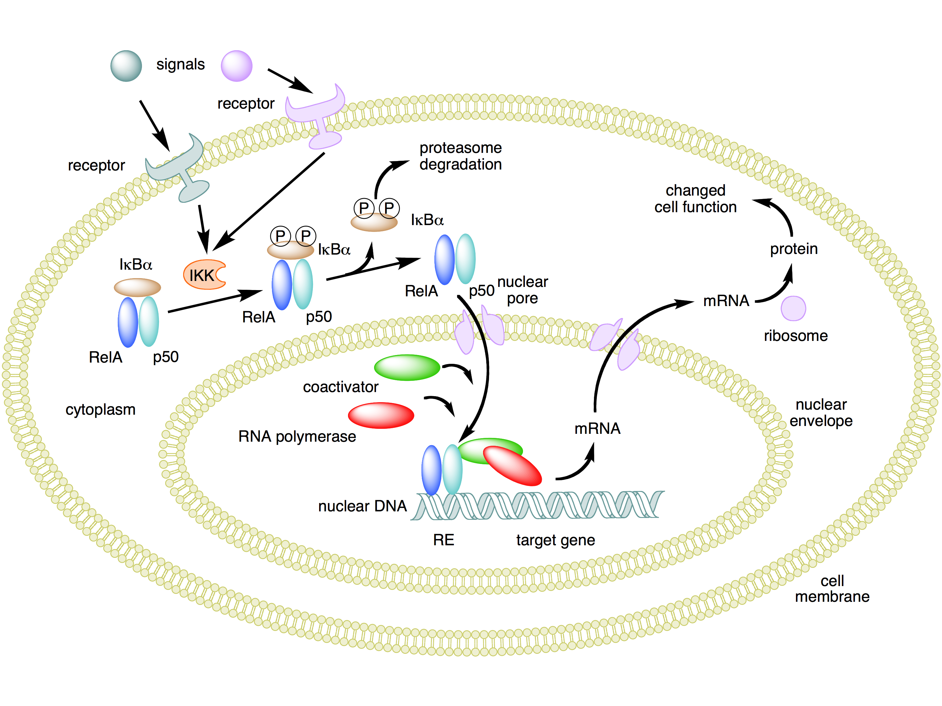
https://upload.wikimedia.org/wikipedia/commons/5/5e/NFKB_mechanism_of_action.png

What Defines a Signaling Pathway Reporter Cell Line?

Transcriptional Coupling of Pathway Activity to Reporter Expression

In these systems, pathway-responsive DNA elements (e.g., response elements or transcription factor binding sites) are placed upstream of a reporter gene. When the signaling pathway is activated, transcription factors bind these elements and induce reporter expression.

An academic overview of transcriptional reporter systems is provided by Cold Spring Harbor Laboratory
https://www.cshl.edu/reporter-genes-and-signaling-pathways/

Common Reporter Genes Used

Typical reporter readouts include:

  • Luciferase (Firefly, NanoLuc) – high sensitivity, wide dynamic range

  • Fluorescent proteins (GFP, mCherry) – live-cell imaging compatibility

  • Secreted reporters (SEAP, Gaussia luciferase) – non-destructive sampling

An NIH-supported review of reporter gene technologies
https://pmc.ncbi.nlm.nih.gov/articles/PMC4553235/

Major Signaling Pathways Studied Using Reporter Cell Lines

NF-κB Pathway Reporter Cell Lines

NF-κB reporters are widely used to study inflammation, immune signaling, and cellular stress responses.

NIH overview of NF-κB signaling
https://www.ncbi.nlm.nih.gov/books/NBK558285/

University-based educational resource (Johns Hopkins University)
https://pathology.jhu.edu/research/research-labs/nf-kb-signaling

MAPK/ERK Pathway Reporter Cell Lines

MAPK reporters enable quantification of growth factor signaling, cell proliferation, and oncogenic pathway activation.

NIH-hosted overview of MAPK signaling
https://www.ncbi.nlm.nih.gov/books/NBK557535/

Educational review from the University of Colorado
https://www.colorado.edu/lab/erksignaling/pathway

JAK/STAT Pathway Reporter Cell Lines

JAK/STAT reporters are commonly applied in cytokine signaling, immune modulation, and transcriptional activation studies.

NIH review of JAK/STAT signaling
https://pmc.ncbi.nlm.nih.gov/articles/PMC7086960/

Educational material from Yale University
https://medicine.yale.edu/immunobiology/research/jak-stat/

Wnt/β-Catenin Pathway Reporter Cell Lines

Wnt reporters are extensively used in developmental biology, stem cell research, and cell fate determination studies.

NIH background on Wnt signaling
https://www.ncbi.nlm.nih.gov/books/NBK546152/

University of Michigan educational resource
https://open.umich.edu/find/open-educational-resources/biology/wnt-signaling

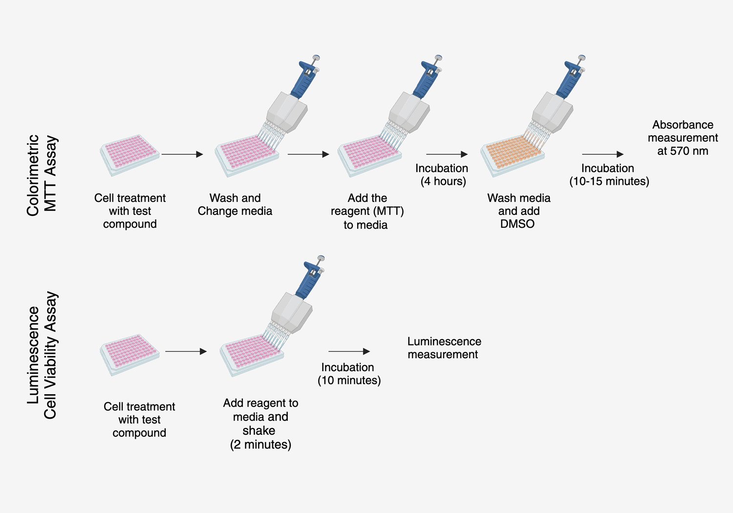
https://byonoy.com/site/assets/files/2908/cellviability_assay-absorbance_vs_luminescence_technology.jpg

Notch, Hedgehog, and TGF-β Pathway Reporters

Reporter cell lines targeting these pathways enable investigation of:

  • Cell differentiation and fate decisions

  • Developmental signaling networks

  • Crosstalk between pathways

NIH overview of Notch signaling
https://www.ncbi.nlm.nih.gov/books/NBK507685/

NIH overview of TGF-β signaling
https://www.ncbi.nlm.nih.gov/books/NBK558366/

Core Laboratory Applications of Signaling Pathway Reporter Cell Lines

1) Mechanism-of-Action Studies

Reporter cell lines provide direct evidence of pathway engagement, allowing researchers to distinguish between on-target and off-target effects.

NIH-supported discussion of pathway-based assays
https://pmc.ncbi.nlm.nih.gov/articles/PMC5742243/

2) High-Throughput Screening (HTS)

Stable reporter cell lines are ideally suited for automation and large-scale screening.

The National Center for Advancing Translational Sciences provides resources on HTS assay development
https://ncats.nih.gov/translation/screening

3) Toxicology and Stress Response Research

Pathway reporters enable sensitive detection of:

  • Oxidative stress responses

  • DNA damage signaling

  • Inflammatory pathway activation

U.S. government background on cellular stress pathways
https://www.ncbi.nlm.nih.gov/books/NBK541154/

Genetic Engineering and Validation of Reporter Cell Lines

Stable Integration Strategies

Reporter constructs are introduced using:

  • Lentiviral or retroviral vectors

  • Antibiotic-based selection systems

  • CRISPR/Cas9-mediated targeted insertion

NIH-funded overview of genome editing strategies
https://pmc.ncbi.nlm.nih.gov/articles/PMC6393419/

https://www.alstembio.com/web/images/services/cell-line-gen/cell_line_generation.png

Signal Validation and Assay Performance

Validated reporter lines are assessed for:

  • Signal-to-background ratio

  • Dose-dependent responsiveness

  • Temporal activation kinetics

University protocol example (University of Wisconsin–Madison)
https://stemcells.wisc.edu/documents/ReporterAssayValidation.pdf

Quality Control, Authentication, and Reproducibility

Long-term experimental reliability depends on identity verification.

Guidance from the National Institute of Standards and Technology
https://www.nist.gov/programs-projects/cell-line-authentication

Supporting background from NCBI Bookshelf
https://www.ncbi.nlm.nih.gov/books/NBK144066/

Comparison: Pathway Reporter Cell Lines vs Biochemical Assays

Feature Reporter Cell Lines Biochemical Assays
Cellular Context Intact Often cell-free
Temporal Resolution High Limited
HTS Compatibility Excellent Variable
Pathway Crosstalk Preserved Reduced

NIH comparison discussion
https://pmc.ncbi.nlm.nih.gov/articles/PMC4553235/

Regulatory and Documentation Awareness (Research Use Only)

For laboratory documentation awareness, U.S. government resources include:

(Provided strictly for research and documentation awareness; not clinical guidance.)

https://www.researchgate.net/publication/335384797/figure/fig1/AS%3A11431281246029581%401716281354427/Workflow-of-cell-line-authentication-by-STR-profiling-The-process-of-the-authentication.png

Why High-Quality Signaling Pathway Reporter Cell Lines Matter

Well-characterized Signaling Pathway Reporter Cell Lines deliver:

  • Quantitative, pathway-specific readouts

  • High reproducibility across experiments

  • Compatibility with multiple detection platforms

  • Scalability for screening and systems biology

They are essential tools for modern signal transduction research and functional cell-based assays.

Signaling Pathway Reporter Cell Lines are powerful research tools that enable real-time, quantitative monitoring of intracellular signaling pathways. Supported by extensive .edu and .gov literature, these stable cellular systems are widely used for pathway analysis, mechanism-of-action studies, and high-throughput screening.

Meta title: Signaling Pathway Reporter Cell Lines for Research | Cell-Based Pathway Analysis
Meta description: Learn how Signaling Pathway Reporter Cell Lines are used in laboratory research for quantitative pathway analysis, functional assays, and high-throughput screening, supported by authoritative .edu and .gov sources.

GPCR Stable Cell Lines in Laboratory Research: Signal Transduction, Functional Assays, and Drug Discovery Models

Introduction: Why GPCR Stable Cell Lines Are Central to Cell-Based Research

G-Protein-Coupled Receptors (GPCRs) represent the largest family of membrane receptors in eukaryotic cells and regulate a vast range of intracellular signaling pathways. GPCR Stable Cell Lines are engineered cell lines that constitutively express a specific GPCR, enabling reproducible, quantitative, and scalable functional assays.

According to the National Institutes of Health, GPCRs mediate cellular responses to hormones, neurotransmitters, and environmental stimuli and are essential for signal transduction research
https://www.ncbi.nlm.nih.gov/books/NBK6285/

In laboratory settings, GPCR stable cell lines are indispensable tools for:

  • Receptor signaling pathway analysis

  • Ligand screening and functional profiling

  • Pharmacological mechanism studies

  • High-throughput screening (HTS) assay development

https://www.abmgood.com/assets/customservice/stable_cell_line_generation/Custom-Stable-Cell-Line-Generation-Service-Workflow-Sept.png

Biological Fundamentals of GPCR Signaling

Canonical GPCR Signal Transduction

GPCRs transmit extracellular signals through heterotrimeric G proteins, activating downstream pathways such as:

  • Gs / Gi → cAMP modulation

  • Gq → intracellular calcium mobilization

  • β-arrestin pathways → receptor desensitization and biased signaling

A detailed educational overview of GPCR signaling is provided by National Center for Biotechnology Information
https://www.ncbi.nlm.nih.gov/books/NBK559299/

An academic signaling overview from Harvard University
https://projects.iq.harvard.edu/files/pharmacology/files/gpcr_signaling.pdf

What Defines a GPCR Stable Cell Line?

GPCR Stable Cell Lines are generated by integrating GPCR-encoding constructs into the host genome, ensuring persistent receptor expression across multiple passages.

Key defining features include:

  • Stable, homogeneous receptor expression

  • Consistent signal amplitude and kinetics

  • Compatibility with multiple assay readouts

A university-level overview of stable cell line generation can be found at Stanford University
https://med.stanford.edu/content/dam/sm/pharmthera/documents/Stable_Cell_Lines.pdf

Core Laboratory Applications of GPCR Stable Cell Lines

1) Ligand Screening and Functional Pharmacology

GPCR stable cell lines enable direct measurement of receptor activation or inhibition in response to ligands.

Common assay formats include:

  • Agonist and antagonist screening

  • Dose–response curve generation

  • Potency (EC₅₀) and efficacy analysis

An NIH-supported overview of GPCR drug screening
https://pmc.ncbi.nlm.nih.gov/articles/PMC5742243/

2) Second Messenger Assays (cAMP and Calcium)

https://media.springernature.com/m685/springer-static/image/art%3A10.1038%2Fnmeth.f.211/MediaObjects/41592_2008_Article_BFnmethf211_Fig1_HTML.jpg

Second messenger measurements remain the gold standard for GPCR functional assays.

  • cAMP assays (Gs/Gi signaling)

  • Calcium flux assays (Gq signaling)

A detailed NIH protocol for calcium signaling assays
https://pubmed.ncbi.nlm.nih.gov/31156626/

Educational assay background from the University of Michigan
https://open.umich.edu/find/open-educational-resources/biology/gpcr-calcium-signaling

3) β-Arrestin Recruitment and Biased Signaling

Modern GPCR research increasingly focuses on biased agonism, where ligands preferentially activate specific pathways.

β-arrestin-based reporter systems enable:

  • Dissection of signaling bias

  • Comparison of ligand functional selectivity

A comprehensive NIH review on biased GPCR signaling
https://pmc.ncbi.nlm.nih.gov/articles/PMC6100574/

Reporter-Based Readouts in GPCR Stable Cell Lines

GPCR stable cell lines are often coupled with constitutive or pathway-specific reporter genes, such as:

  • Luciferase (CRE, SRE, NFAT reporters)

  • Fluorescent protein reporters

  • Enzyme-based readouts

An NIH-indexed overview of reporter gene assays
https://pmc.ncbi.nlm.nih.gov/articles/PMC4553235/

High-Throughput Screening (HTS) and Automation Compatibility

Because of their robustness and signal consistency, GPCR stable cell lines are widely used in automated screening environments.

The National Center for Advancing Translational Sciences provides educational resources on HTS assay development
https://ncats.nih.gov/translation/screening

A university guide to HTS assay optimization
https://med.stanford.edu/cvi/research/hts.html

Genetic Engineering and Quality Control Considerations

Integration and Expression Stability

GPCR genes are commonly introduced via:

  • Lentiviral transduction

  • Retroviral systems

  • Targeted genome integration

An NIH-funded overview of genome engineering strategies
https://pmc.ncbi.nlm.nih.gov/articles/PMC6393419/

Cell Line Authentication and Reproducibility

https://www.researchgate.net/publication/335384797/figure/fig1/AS%3A11431281246029581%401716281354427/Workflow-of-cell-line-authentication-by-STR-profiling-The-process-of-the-authentication.png

Long-term reproducibility depends on identity verification and contamination control.

Guidelines from the National Institute of Standards and Technology
https://www.nist.gov/programs-projects/cell-line-authentication

Supporting background from NCBI Bookshelf
https://www.ncbi.nlm.nih.gov/books/NBK144066/

Comparison: GPCR Stable Cell Lines vs Transient Expression Systems

Feature GPCR Stable Cell Lines Transient Transfection
Expression Consistent Variable
Reproducibility High Moderate
HTS Suitability Excellent Limited
Setup Time Longer Shorter
Assay Robustness High Variable

A comparative academic discussion is available via PubMed Central
https://pmc.ncbi.nlm.nih.gov/articles/PMC5742243/

Regulatory and Documentation Awareness (Research Use Only)

For laboratory documentation and biosafety awareness, U.S. government resources include:

(Provided strictly for research and documentation awareness; not clinical guidance.)

Why High-Quality GPCR Stable Cell Lines Matter in Research

Well-characterized GPCR Stable Cell Lines provide:

  • Predictable and reproducible receptor signaling

  • High signal-to-noise ratios

  • Compatibility with multiple readout technologies

  • Scalability for screening and mechanistic studies

They are foundational tools for quantitative GPCR biology, pharmacology, and cell-based assay development.

GPCR Stable Cell Lines are essential research tools that enable robust analysis of receptor-mediated signaling, ligand pharmacology, and pathway-specific responses. Supported by extensive .edu and .gov literature, these stable cellular models are widely used in functional assays, reporter-based readouts, and high-throughput screening workflows.

Meta title: GPCR Stable Cell Lines for Research | Cell-Based Assays, Signaling & Drug Screening
Meta description: Explore how GPCR Stable Cell Lines are used in laboratory research for receptor signaling analysis, ligand screening, second-messenger assays, and high-throughput screening, supported by authoritative .edu and .gov sources.

Constitutive Reporter Cell Lines in Laboratory Research: Stable Signal Expression, Assay Development, and Quantitative Readouts

Introduction: Why Constitutive Reporter Cell Lines Are Essential Experimental Tools

Constitutive Reporter Cell Lines are genetically engineered cell lines that express a reporter gene continuously and stably, under the control of a constitutive promoter. Unlike inducible systems, constitutive reporters provide a constant, measurable signal that reflects cell number, viability, transcriptional activity, or pathway integrity.

According to the National Center for Biotechnology Information, reporter genes encode proteins that can be easily measured to study cellular processes
https://www.ncbi.nlm.nih.gov/books/NBK6360/

In modern laboratories, constitutive reporter cell lines are foundational tools for:

  • Cell-based assay development

  • High-throughput screening (HTS)

  • Signal normalization and internal controls

  • Cytotoxicity and proliferation studies

  • Transfection efficiency benchmarking

What Defines a Constitutive Reporter Cell Line?

Stable Reporter Expression Independent of Experimental Stimuli

In constitutive systems, reporter genes are driven by ubiquitously active promoters such as CMV, EF1α, or PGK, ensuring signal stability across experimental conditions.

A university-level overview of constitutive promoter usage is provided by Addgene (Harvard-affiliated)
https://www.addgene.org/guides/promoters/

Common Reporter Genes Used

Widely used reporter genes include:

  • Luciferase (Firefly, Renilla, NanoLuc)

  • Green Fluorescent Protein (GFP) and variants

  • β-galactosidase (LacZ)

  • Secreted alkaline phosphatase (SEAP)

An NIH-supported review of reporter gene technologies is available via PubMed Central
https://pmc.ncbi.nlm.nih.gov/articles/PMC4553235/

Core Applications of Constitutive Reporter Cell Lines

https://www.cell.com/cms/10.1016/j.isci.2023.106156/asset/d8db7c1c-2d83-4845-968f-6d1c3bc7caa1/main.assets/fx1_lrg.jpg

1) Cell Viability and Proliferation Assays

Because reporter signal correlates directly with viable cell number, constitutive reporters are widely used for:

  • Cytotoxicity testing

  • Drug sensitivity profiling

  • Growth curve quantification

An academic example of luciferase-based viability assays is described by the National Institutes of Health
https://pubmed.ncbi.nlm.nih.gov/28719231/

2) Normalization Controls in Cell-Based Assays

Constitutive reporters often serve as internal normalization controls, allowing researchers to:

  • Correct for cell number variation

  • Reduce inter-well variability

  • Improve assay robustness

This approach is discussed in a methods review from Stanford University
https://med.stanford.edu/content/dam/sm/cvi/documents/Reporter_Assays_Review.pdf

3) High-Throughput Screening (HTS)

Stable reporter cell lines are highly compatible with automated screening platforms, making them indispensable in:

  • Compound libraries screening

  • Toxicology research

  • Functional genomics

The National Center for Advancing Translational Sciences provides background on HTS assay development
https://ncats.nih.gov/translation/screening

Genetic Engineering and Generation of Stable Reporter Lines

https://www.abmgood.com/assets/customservice/stable_cell_line_generation/Custom-Stable-Cell-Line-Generation-Service-Workflow-Sept.png

Integration Strategies

Reporter constructs are commonly introduced via:

  • Lentiviral or retroviral transduction

  • Plasmid transfection followed by selection

  • CRISPR/Cas9-mediated knock-in

An NIH-funded overview of genome editing strategies in cell lines
https://pmc.ncbi.nlm.nih.gov/articles/PMC6393419/

Selection and Validation

After integration, stable cell lines are selected using antibiotics (e.g., puromycin, neomycin) and validated for:

  • Signal stability across passages

  • Signal-to-background ratio

  • Linear response to cell number

A university protocol example (University of Wisconsin–Madison)
https://stemcells.wisc.edu/documents/StableCellLineGeneration.pdf

Reporter Detection Technologies and Readout Platforms

Luminescence-Based Reporters

Luciferase reporters offer:

  • High sensitivity

  • Wide dynamic range

  • Low background

NIH-hosted comparison of luminescent reporters
https://pubmed.ncbi.nlm.nih.gov/30388435/

Fluorescence-Based Reporters

Fluorescent reporters enable:

  • Live-cell imaging

  • Spatial analysis

  • Flow cytometry compatibility

A comprehensive educational resource from Cold Spring Harbor Laboratory
https://cshl.edu/reporter-genes-fluorescent-proteins/

Quality Control, Stability, and Reproducibility

https://www.researchgate.net/publication/335384797/figure/fig1/AS%3A11431281246029581%401716281354427/Workflow-of-cell-line-authentication-by-STR-profiling-The-process-of-the-authentication.png

Genetic and Identity Verification

Long-term reporter stability depends on rigorous identity control.

Guidance from the National Institute of Standards and Technology
https://www.nist.gov/programs-projects/cell-line-authentication

Additional authentication background from NCBI Bookshelf
https://www.ncbi.nlm.nih.gov/books/NBK144066/

Passage-Dependent Signal Monitoring

Best practices include:

  • Monitoring reporter intensity across passages

  • Avoiding over-confluence

  • Freezing early-passage master stocks

Comparison: Constitutive vs Inducible Reporter Systems

Feature Constitutive Reporters Inducible Reporters
Signal Continuous Stimulus-dependent
Complexity Low Higher
Variability Minimal Context-dependent
Best Use Controls, HTS, viability Pathway activation

A detailed academic comparison is available via PubMed Central
https://pmc.ncbi.nlm.nih.gov/articles/PMC5761588/

Regulatory and Documentation Awareness (Research Use Only)

For laboratory documentation and compliance awareness, U.S. government resources include:

(Provided for research documentation awareness only; not clinical guidance.)

Why High-Quality Constitutive Reporter Cell Lines Matter

Well-characterized Constitutive Reporter Cell Lines provide:

  • Stable and reproducible signals

  • Reduced assay variability

  • Improved data normalization

  • Compatibility with automation and HTS

They are essential tools for quantitative, scalable, and reproducible cell-based research.

Constitutive Reporter Cell Lines are indispensable research tools that enable stable, continuous reporter gene expression for cell-based assays, high-throughput screening, cytotoxicity testing, and signal normalization. Supported by extensive .edu and .gov literature, these systems provide reliable quantitative readouts across diverse experimental platforms.

Meta title: Constitutive Reporter Cell Lines for Research | Stable Reporter Expression & Cell-Based Assays
Meta description: Discover how Constitutive Reporter Cell Lines are used in laboratory research for viability assays, normalization controls, and high-throughput screening, supported by authoritative .edu and .gov references.

Embryonic Stem Cells (ESCs) in Laboratory Research: Pluripotency, Differentiation Models, and Advanced Experimental Applications

Introduction: Embryonic Stem Cells as a Gold Standard for Pluripotency Research

Embryonic Stem Cells (ESCs) are pluripotent cells derived from the inner cell mass of the blastocyst-stage embryo. In laboratory research, ESCs represent the reference model for pluripotency, offering unparalleled capacity to differentiate into derivatives of all three germ layers: ectoderm, mesoderm, and endoderm.

According to the National Institutes of Health, embryonic stem cells are defined by their unlimited self-renewal capacity and pluripotent differentiation potential
https://stemcells.nih.gov/info/basics/stc-basics

ESCs are widely used in basic and translational research to study early developmental biology, gene regulation, lineage specification, and disease-relevant cellular phenotypes—strictly within research-use-only laboratory frameworks.

AffiCELL® ICR Mouse Embryonic Fibroblasts (Inactivated)

Core Biological Properties of Embryonic Stem Cells

True Pluripotency and Unlimited Self-Renewal

Unlike multipotent adult stem cells, ESCs maintain:

  • Stable pluripotent transcriptional programs

  • Long-term proliferative capacity

  • Consistent differentiation responsiveness

A comprehensive academic review on ESC pluripotency is provided by Stanford University
https://med.stanford.edu/content/dam/sm/stemcell/documents/publications/StemCells_ESC_review.pdf

Open Chromatin and Epigenetic Plasticity

ESCs exhibit a highly permissive chromatin state, making them ideal for:

  • Epigenetic regulation studies

  • Chromatin remodeling research

  • Developmental gene network mapping

An in-depth NIH-supported discussion of epigenetic regulation in ESCs can be found here
https://pmc.ncbi.nlm.nih.gov/articles/PMC3574585/ESC Pluripotency Markers and Identity Verification

Canonical Pluripotency Marker Expression

ESC identity is commonly verified using transcription factors such as:

  • OCT4 (POU5F1)

  • SOX2

  • NANOG

Marker expression and maintenance are extensively discussed by Harvard University
https://stemcell.harvard.edu/science/escs

Quality Control and Cell Authentication

Long-term ESC culture requires stringent quality control to prevent:

  • Spontaneous differentiation

  • Genetic drift

  • Cross-contamination

Guidelines for cell identity and authentication are outlined by the National Institute of Standards and Technology
https://www.nist.gov/programs-projects/cell-line-authentication

Additional reference from the National Center for Biotechnology Information
https://www.ncbi.nlm.nih.gov/books/NBK144066/

https://www.researchgate.net/publication/8551939/figure/fig3/AS%3A277679652786199%401443215397057/Differentiation-to-all-three-embryonic-germ-layers-and-subsequently-to-different-tissues.png

Directed Differentiation of ESCs into Germ Layer Lineages

ESCs serve as a foundational model for directed differentiation protocols, enabling controlled generation of lineage-specific progenitors.

Common Laboratory Differentiation Models

  • Ectoderm: neural progenitors, sensory lineages

  • Mesoderm: cardiomyocytes, hematopoietic progenitors

  • Endoderm: hepatic and pancreatic progenitors

An NIH-funded overview of germ layer differentiation strategies is available at
https://pmc.ncbi.nlm.nih.gov/articles/PMC5903004/

A university-based differentiation protocol example (University of Wisconsin–Madison)
https://stemcells.wisc.edu/research/embryonic-stem-cells/

ESCs in Developmental Biology and Early Patterning Studies

ESCs uniquely enable in vitro modeling of early embryogenesis, including:

  • Axis formation

  • Morphogen signaling gradients

  • Cell fate decisions

A detailed academic discussion of early developmental modeling is provided by Princeton University
https://molbio.princeton.edu/sites/default/files/ESC_Development_Review.pdf

Genetic Engineering and Functional Genomics in ESCs

ESCs are highly amenable to:

  • CRISPR/Cas9 genome editing

  • Reporter gene insertion

  • Loss- and gain-of-function studies

Because of their stable self-renewal, ESCs are often used for developmental gene regulatory network analysis.

NIH-supported guidance on genome editing in pluripotent stem cells
https://pmc.ncbi.nlm.nih.gov/articles/PMC6393419/

https://www.researchgate.net/publication/294872498/figure/fig49/AS%3A330143458447374%401455723742900/ESC-differentiation-into-ectodermal-mesodermal-and-endodermal-derivatives-Wnt-BMP-and.png

Omics-Based Discovery Using Embryonic Stem Cells

ESCs are widely employed in:

  • Bulk and single-cell RNA sequencing

  • Chromatin accessibility (ATAC-seq)

  • Histone modification mapping (ChIP-seq)

A systems-level overview of pluripotent stem cell omics is available via PubMed Central
https://pmc.ncbi.nlm.nih.gov/articles/PMC6364749/

Ethical Oversight and Regulatory Awareness (Research Context Only)

All ESC research is conducted under strict ethical and institutional oversight. In the United States, federally funded ESC research follows clearly defined policy frameworks.

Key authoritative resources include:

(Provided strictly for research documentation awareness; not clinical guidance.)

Why High-Quality Embryonic Stem Cell Products Matter in Research

When selecting Embryonic Stem Cells for laboratory research, scientists typically prioritize:

  • Verified pluripotency marker expression

  • Genetic stability and passage consistency

  • Responsiveness to differentiation protocols

  • Clear research-use-only documentation

High-quality ESC products support robust developmental models, reproducible differentiation, and high-impact academic research.

Embryonic Stem Cells (ESCs) are the gold-standard pluripotent model for laboratory research, enabling in-depth studies of early development, germ layer differentiation, epigenetic regulation, and genome-wide functional analysis. Supported by extensive .edu and .gov literature, ESC-based systems remain essential tools in developmental and molecular biology laboratories worldwide.

Mesenchymal Stem Cells (MSCs) in Laboratory Research: Advanced Applications, Experimental Models, and Best Practices

Introduction: Why Mesenchymal Stem Cells Remain Central to Experimental Biology

Mesenchymal Stem Cells (MSCs)—also referred to as mesenchymal stromal cells—are among the most widely studied cell types in basic, translational, and preclinical laboratory research. Their ability to self-renew, differentiate into multiple mesodermal lineages, and interact dynamically with surrounding cells makes them a foundational model system across numerous biological disciplines.

According to the National Cancer Institute, mesenchymal stem cells are defined as multipotent stromal cells capable of differentiating into osteoblasts, adipocytes, and chondrocytes
(https://www.cancer.gov/publications/dictionaries/cancer-drug/def/mesenchymal-stem-cell).

In laboratory settings, MSCs are not used as therapeutic agents, but rather as highly adaptable research tools that enable mechanistic studies in:

  • Cell differentiation and lineage commitment

  • Immune cell interaction and signaling

  • Tissue remodeling and matrix biology

  • Extracellular vesicle (EV) and exosome research

  • Transcriptomic, epigenomic, and proteomic discovery

AffiCELL® Wistar Rat Adipose-derived Mesenchymal Stem Cells

Biological Characteristics That Make MSCs Ideal for Research Models

Plastic Adherence and In-Vitro Stability

MSCs readily adhere to standard tissue-culture plastic, allowing robust expansion under defined conditions. This practical feature supports longitudinal experiments, multi-passage studies, and comparative assays.

The National Institutes of Health provides extensive background on stromal cell culture systems via PubMed Central
https://pmc.ncbi.nlm.nih.gov/articles/PMC6431372/

Multipotent Differentiation Capacity

Under lineage-specific induction conditions, MSCs differentiate into:

  • Osteogenic lineage (mineralization, alkaline phosphatase activity)

  • Adipogenic lineage (lipid droplet accumulation)

  • Chondrogenic lineage (proteoglycan and collagen II expression)

A comprehensive academic overview of MSC differentiation assays is available through the University of North Carolina
https://bme.unc.edu/wp-content/uploads/sites/917/2022/04/Stem-Cells-2014-Lv.pdf

MSC Phenotyping and Identity Verification in the Laboratory

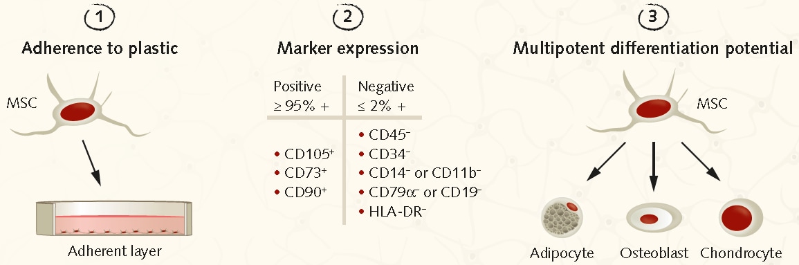
https://www.sigmaaldrich.com/deepweb/assets/sigmaaldrich/marketing/global/images/technical-documents/articles/cell-culture-and-analysis/imaging-analysis-and-live-cell-imaging/minimum-isct-criteria/minimum-isct-criteria.png

Surface Marker Profiling

Flow cytometry remains a cornerstone technique for MSC characterization. Commonly evaluated markers include:

  • Positive expression: CD73, CD90, CD105

  • Negative expression: hematopoietic and endothelial markers

An academic discussion on MSC marker variability can be found at the University of California, San Diego
https://muscle.ucsd.edu/pubs/pdf/Ruoss_AJSM_2021.pdf

Cell Identity and Authentication

While MSCs are often primary-like cultures, identity control remains critical, particularly in long-term studies.

The National Institute of Standards and Technology outlines best practices for human cell identity control
https://www.nist.gov/programs-projects/cell-line-authentication/cell-line-id-and-authentication-human-cell-lines

Additional guidance from the National Center for Biotechnology Information
https://www.ncbi.nlm.nih.gov/books/NBK144066/

MSC Co-Culture Systems and Immunology Research

MSCs are frequently used in co-culture models to study:

  • Cell–cell contact signaling

  • Paracrine communication

  • Cytokine and chemokine regulation

These systems are widely applied in immunology, inflammation biology, and tissue microenvironment modeling.

A peer-reviewed overview from PubMed discusses MSC-mediated immune signaling mechanisms
https://pubmed.ncbi.nlm.nih.gov/32709406/

MSC Secretome, Extracellular Vesicles, and Exosome Research

One of the fastest-growing MSC research areas is the study of the MSC secretome, including extracellular vesicles (EVs) and exosomes.

Laboratory applications include:

  • Conditioned media signaling studies

  • EV-mediated protein and RNA transport

  • Biomarker discovery

Widely cited protocols and methodological references include:

Tissue Engineering and Biomaterials Research Using MSCs

MSCs are extensively used in biomaterials and tissue engineering laboratories to evaluate:

  • Scaffold composition and stiffness

  • Surface chemistry and cell adhesion

  • Mechanical cues influencing lineage fate

The U.S. National Library of Medicine hosts multiple open-access reviews on MSC-based tissue models
https://pubmed.ncbi.nlm.nih.gov/31815001/

Omics and Systems Biology Applications

Because MSCs respond dynamically to environmental cues, they are ideal for:

  • RNA sequencing (bulk and single-cell)

  • Epigenomic profiling (ATAC-seq, ChIP-seq)

  • Proteomics and phosphoproteomics

A comprehensive review of MSC molecular profiling is available via PubMed Central
https://pmc.ncbi.nlm.nih.gov/articles/PMC6364749/

https://prod-shared-star-protocols.s3.amazonaws.com/protocols/3412-GA.jpg

Regulatory and Documentation Awareness (Research Context Only)

For laboratories maintaining structured documentation, regulatory frameworks are often cited for awareness only, not for clinical intent.

Key U.S. government references include:

Why High-Quality MSC Products Matter in Research

When selecting Mesenchymal Stem Cells for laboratory use, researchers typically evaluate:

  • Documented origin and handling conditions

  • Reproducible growth and morphology

  • Compatibility with differentiation, co-culture, and EV workflows

  • Clear research-use-only positioning

Well-characterized MSC products support reproducibility, inter-lab consistency, and high-impact publication outcomes.

Mesenchymal Stem Cells (MSCs) are indispensable tools in modern biological research, enabling differentiation studies, immune interaction modeling, extracellular vesicle analysis, tissue engineering, and multi-omics discovery. Supported by extensive .edu and .gov literature, MSC-based experiments continue to advance our understanding of cellular communication and functional biology in controlled laboratory environments.